Wavelength ranges: VIS, NIR, SWIR
A strip in the camera’s field of view is illuminated and an imaging spectrograph produces sequentially high-resolution spectral and spatial images through its sensors. This technique is also known as push-broom technology. Figure 1 illustrates how it works.
How it works
Technical points
Due to their significant effect on performance, some technical points are worth mentioning:
Short focal length, corrected NIR lenses:
large field of view
ZEISS optics & Offner spectrograph:
high-quality spectra
High-performance image sensor:
Optimised electronics
measurement of extremely fast objects, high throughput
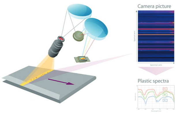
Figure 1: Principle of the Offner setup and data processing
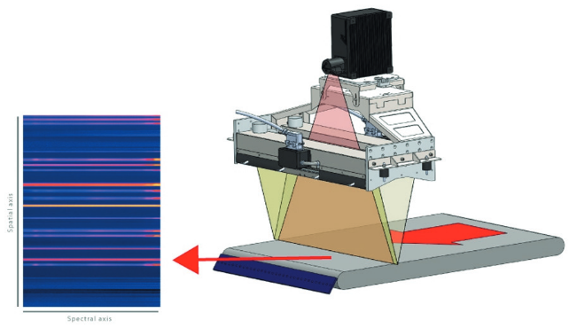
Figure 2: Principle of our camera in operation
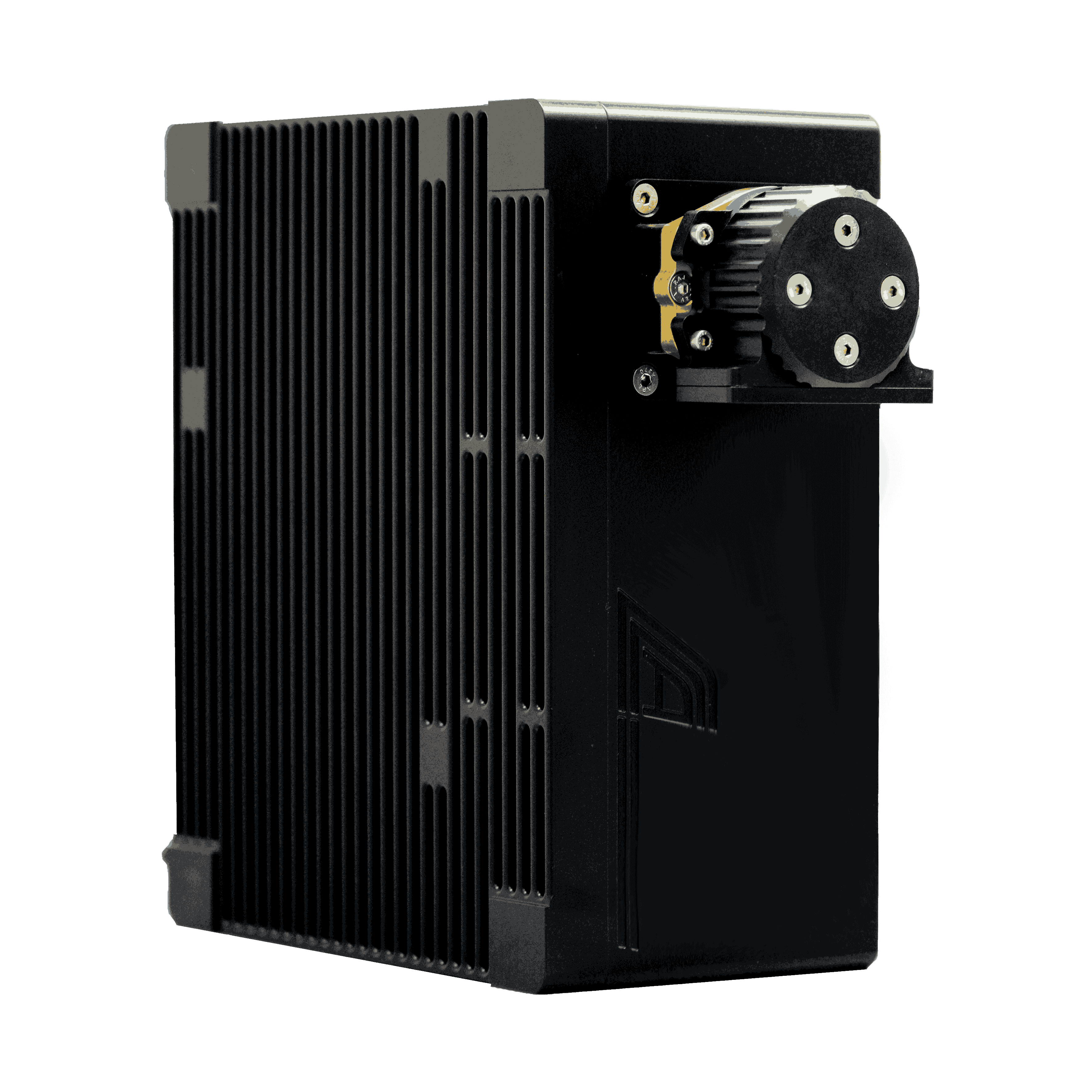
Hyperspectral cameras
Wavelength ranges: VIS/NIR/SWIR
A strip in the camera’s field of view is illuminated and sequentially imaged onto optical fibres arranged in a row. This technique is called push-broom technology. Figure 1 illustrates how it works.
How it works
Technical points
Independent optical fibres:
simultaneous measurement on multiple conveyors
ZEISS optics:
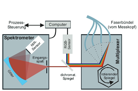
Figure 1: Beam path in multiplexer
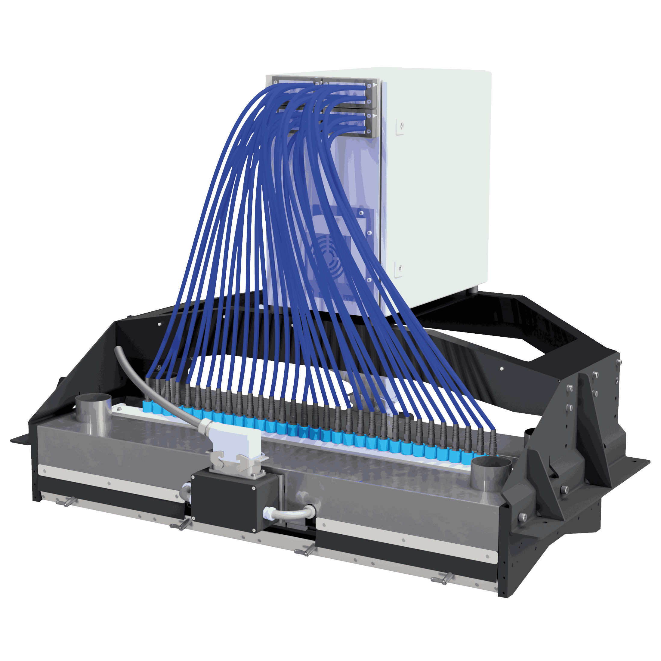
Multiplexed NIR Spectrometer
Wavelength ranges: VIS
A strip in the camera’s field of view is illuminated and sequentially measured at a high frame rate. This technique is called push-broom technology. The beam path is shown in Figure 1 to illustrate how it works.
How it works
Technical points
Optimised electronics:
measurement of extremely fast objects, high throughput
CMOS sensor & ZEISS optics:
high image contrast, low noise
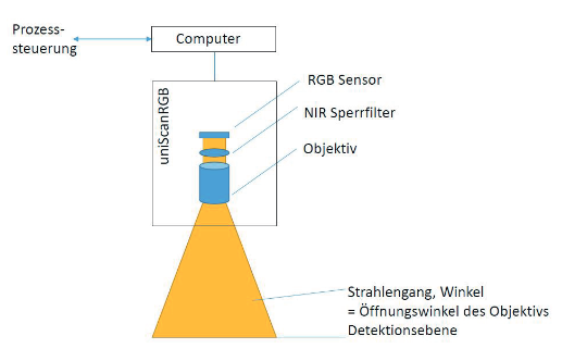
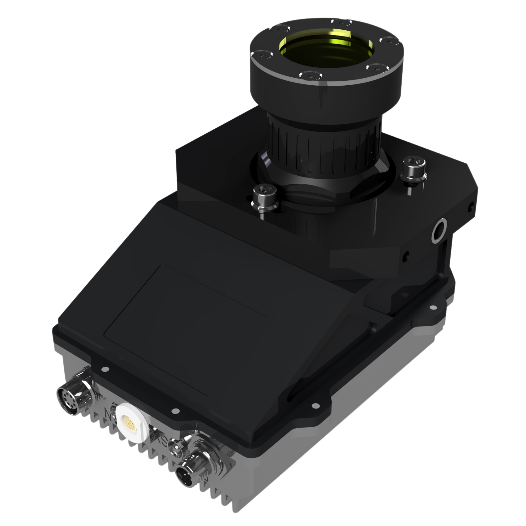
Universal line scan colour camera
Wavelength ranges: X-ray/UV
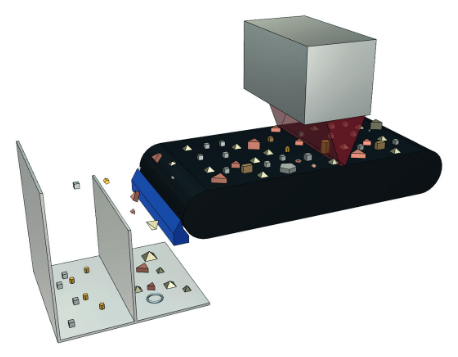
A strip in the detector’s field of view is illuminated and sequentially measured. This technique is also known as push-broom technology.
How it works
Technical points
Water-cooled, high power x-ray tube:
high primary radiation intensity produces high fluorescence radiation intensity
Silicon drift detectors or the latest generation:
high count rates and best-possible energy resolution
Complete system (tube, detector & electronics:
fast and detailed identification of alloys, not only the main elements
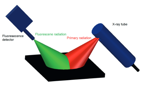
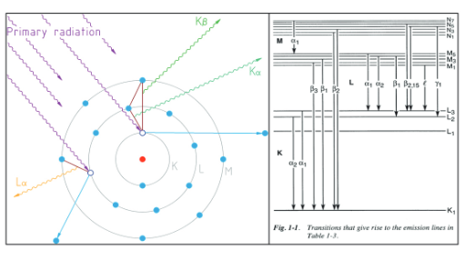
Figure 1: Functionality of x-ray fluorescence analysis
X-ray fluorescence analyser
LLA Instruments GmbH
Justus-von-Liebig Straße 9/11
12489 Berlin
Germany

Justus-von-Liebig
Straße 9/11
12489 Berlin
Germany